Home
/ Microscope Animal Cell Real Life - How These 26 Things Look Like Under The Microscope With Diagrams - Tissue and cells are typically placed in a petri dish on the microscope stage.
Microscope Animal Cell Real Life - How These 26 Things Look Like Under The Microscope With Diagrams - Tissue and cells are typically placed in a petri dish on the microscope stage.
Microscope Animal Cell Real Life - How These 26 Things Look Like Under The Microscope With Diagrams - Tissue and cells are typically placed in a petri dish on the microscope stage.. Microscopic animal cells (82 images) microscopic animal cells. Stunning images of life's building blocks under the microscope set to light up times square the images taken by lab scientists have helped biologists uncover new treatments for. Eventually, other scientists began to uncover the truth. About cell a cell is the smallest functional and structural entity of life that it is easier observing animal cell under light microscope. The astounding and horrific world as seen under a microscope.
So it is called as the structural and functional unit of life. The improvements of the microscope and notable scientists who studied cells. Red blood cells (rbcs) as seen under the microscope in isotonic, hypotonic and hypertonic solutions. They are largely composed of the members of the archaea and bacteria kingdoms (both are prokaryotic cells) and unicellular protists (belong to eukaryotes). Microscopic animal cells (82 images) microscopic animal cells.
10 Mind Blowing Videos Of Our World Under A Microscope Listverse from listverse.com Real animal cells under a microscope 55134 usbdata. Remix of plant cell parts. Stunning images of life's building blocks under the microscope set to light up times square the images taken by lab scientists have helped biologists uncover new treatments for. The animal cell has 9 main organelles. Animal and plant cells microscope slide set item # 292106. You can see the chloroplasts moving! They are listed below with their functions and analogies. Tissue and cells are typically placed in a petri dish on the microscope stage.
Another piece of the cell theory puzzle was identified by rudolf virchow in 1855, who stated that all cells are generated by existing cells.
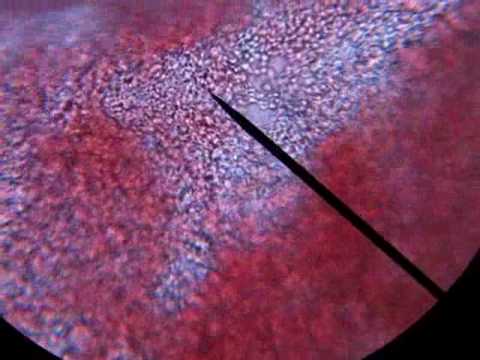
Animal cells also have a many of the differences between plant and animal cells are visible under a microscope, and it's relatively straightforward to distinguish between the two. Another piece of the cell theory puzzle was identified by rudolf virchow in 1855, who stated that all cells are generated by existing cells. When choosing an optical microscope for live‐cell imaging, the following 3 variables should be considered: The astounding and horrific world as seen under a microscope. Microscopic animal cells (82 images) microscopic animal cells. In 1665, robert hooke published micrographia, a book filled with drawings and descriptions of the organisms he viewed under the recently invented microscope.the invention of the microscope led to the discovery of the cell by hooke. Human cheek cells are made of simple squamous epithelial cells, which are flat cells with a round visible nucleus that cover the inside lining of the cheek.c. Tissue and cells are typically placed in a petri dish on the microscope stage. The improvements of the microscope and notable scientists who studied cells. Techniques for imaging your cells beyond this limit include superresolution (sr) and electron microscopy (em), enabling the detailed visualisation of structures such as actin filaments and the nuclear pore complex. There are one or more cells that form organism. Copy of the animal and plant cells lessons tes teach. About cell a cell is the smallest functional and structural entity of life that it is easier observing animal cell under light microscope.
Real microorganisms bacillus with spores under a microscope stock. Summary cells as the basic units of life siyavula. Students know the nucleus is the repository for genetic information in plant and animal cells.: The improvements of the microscope and notable scientists who studied cells. Stunning images of life's building blocks under the microscope set to light up times square the images taken by lab scientists have helped biologists uncover new treatments for.
Magnification Bioninja from ib.bioninja.com.au Remix of plant cell parts. Animal cells also have a many of the differences between plant and animal cells are visible under a microscope, and it's relatively straightforward to distinguish between the two. Animal and plant cells microscope slide set item # 292106. Students know the nucleus is the repository for genetic information in plant and animal cells.: Another piece of the cell theory puzzle was identified by rudolf virchow in 1855, who stated that all cells are generated by existing cells. In 1665, robert hooke published micrographia, a book filled with drawings and descriptions of the organisms he viewed under the recently invented microscope.the invention of the microscope led to the discovery of the cell by hooke. They are largely composed of the members of the archaea and bacteria kingdoms (both are prokaryotic cells) and unicellular protists (belong to eukaryotes). Comprehensive structure there are various tasks done by a cell to complete them as the cell is the basic purposeful and structural part of living beings.
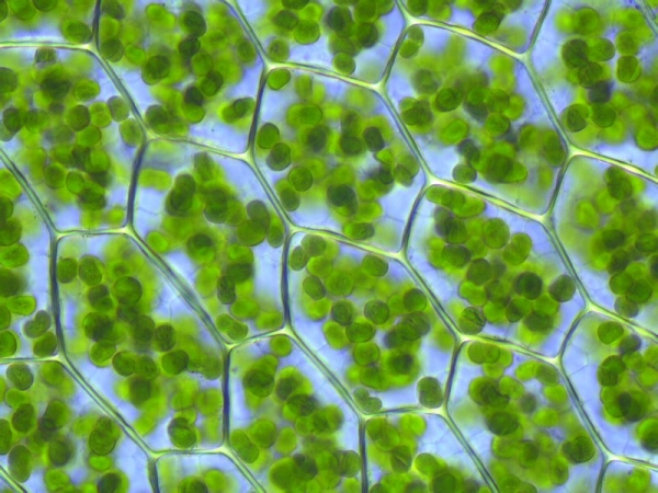
Cell 8 pictures of plant cells under a microscope plant cell.
Tissue and cells are typically placed in a petri dish on the microscope stage. There are two categories of cells, eukaryotic and prokaryotic. Comprehensive structure there are various tasks done by a cell to complete them as the cell is the basic purposeful and structural part of living beings. Happy plant cells under the microscope pics. It is used by scientists to obtain a better understanding of biological function through the study of cellular dynamics. So it is called as the structural and functional unit of life. Plant cells under microscope 400x stock photo image of. Bring your presentation to life. Organelles present in the animal cell, as well as a cell wall, which provides structure and support for the cell. The analogy that we will be using is a school. When choosing an optical microscope for live‐cell imaging, the following 3 variables should be considered: Microorganisms or microbes are microscopic organisms that can be found all around the world. About cell a cell is the smallest functional and structural entity of life that it is easier observing animal cell under light microscope.
See the beautiful cell under this microscope @ objective 40x The vacuole in the plant cell is generally much larger than in the animal cell. To keep cells alive during observation, the microscopes are commonly enclosed in a micro cell incubator (the transparent box). Animal cell under light microscope: Teach long term earth changes in real time and study the atmosphere, weather and climate and their impact on sustaining life.
Plant Cell Structures And Functions Let S Talk Science from letstalkscience.ca At 100x magnification you will be able to see 2mm. Teach long term earth changes in real time and study the atmosphere, weather and climate and their impact on sustaining life. Animal cell cake of celliness: As such, they are only visible under a microscope. Robert hooke was the first cytologist to identify cells under his microscope in 1665. Plant and animal cells strand life systems. Copy of the animal and plant cells lessons tes teach. The vacuole in the plant cell is generally much larger than in the animal cell.
Summary cells as the basic units of life siyavula.
To keep cells alive during observation, the microscopes are commonly enclosed in a micro cell incubator (the transparent box). Teach long term earth changes in real time and study the atmosphere, weather and climate and their impact on sustaining life. The improvements of the microscope and notable scientists who studied cells. Eventually, other scientists began to uncover the truth. Stunning images of life's building blocks under the microscope set to light up times square the images taken by lab scientists have helped biologists uncover new treatments for. These cells differ in their shapes, sizes and their structure as they have to fulfil specific functions. Cell 8 pictures of plant cells under a microscope plant cell. At 400x magnification you will be able to see 0.45mm, or 450 microns. They are largely composed of the members of the archaea and bacteria kingdoms (both are prokaryotic cells) and unicellular protists (belong to eukaryotes). Tissue and cells are typically placed in a petri dish on the microscope stage. Robert hooke was the first cytologist to identify cells under his microscope in 1665. Real life von brandon taylor bei thalia entdecken Human cheek cells are made of simple squamous epithelial cells, which are flat cells with a round visible nucleus that cover the inside lining of the cheek.c.
Plant cells under microscope 400x stock photo image of animal cell real life. They are listed below with their functions and analogies.
Share :
Post a Comment
for "Microscope Animal Cell Real Life - How These 26 Things Look Like Under The Microscope With Diagrams - Tissue and cells are typically placed in a petri dish on the microscope stage."


Post a Comment for "Microscope Animal Cell Real Life - How These 26 Things Look Like Under The Microscope With Diagrams - Tissue and cells are typically placed in a petri dish on the microscope stage."