Home
/ Animal Cell Labeled With Cytoskeleton / Eukaryotic Cells Biology I / A structural scaffold giving the cell shape, an intracellular transport system, a driver of cell motility, and a the cytoskeleton is a dynamic 3d protein network connected to the membrane and some organelles.
Animal Cell Labeled With Cytoskeleton / Eukaryotic Cells Biology I / A structural scaffold giving the cell shape, an intracellular transport system, a driver of cell motility, and a the cytoskeleton is a dynamic 3d protein network connected to the membrane and some organelles.
Animal Cell Labeled With Cytoskeleton / Eukaryotic Cells Biology I / A structural scaffold giving the cell shape, an intracellular transport system, a driver of cell motility, and a the cytoskeleton is a dynamic 3d protein network connected to the membrane and some organelles.. The cytoskeleton also forms tracks. The cytoskeleton, depending on the cell type, is assembled from one or more of three major structural fibers: The function of the cell wall or cytoskeleton is to keep the cell in its shape and to keep it from squishing. After fixation and labelling with specific probes. The cell cytoskeleton serves to protect the cell from both pulling (tensile) and pushing (compression) stress, so maintaining the cell tensegrity.
After fixation and labelling with specific probes. Most of the cells size range between 1 and 100 micrometers and are visible only with the microscope. Eukaryotic cells are larger, more complex, and have evolved more recently than prokaryotes. Animal cells as seen in the fluorescence microscope. The actin cytoskeleton was visualized using alexa fluor 647 phalloidin and alexa fluor 488 phalloidin (cell signaling technology, leiden, the netherlands), respectively.
Bildungsdiagramm Der Biologie Fur Tierzelldiagramm Lizenzfrei Nutzbare Vektorgrafiken Clip Arts Illustrationen Image 80712824 from previews.123rf.com The cytoskeleton also provides cellular stability and controls cell movement (flagella, etc). Unlike the eukaryotic cells of plants and fungi, animal cells do not have a cell wall. Plant cells display a singular architecture, necessitating a structurally and functionally unique cytoskeleton and plant specific control mechanisms. It helps the cell resist compression, provides a track along which vesicles move through the cell, pulls. They are stained with fluorescent labels to help visualise the cytoskeleton with microtubules (green), actin cells would not be cells with out their cytoskeleton (images courtesy of mark shipman, james blyth and louise cramer, laboratory for molecular cell. Where, prokaryotes are just bacteria and archaea to check if you have understood the cell parts, draw a blank animal cell diagram and try to fill in the different parts without referring to the labeled one given. Of the many proteins that interact with it fig. The inner part of the cell and which, with the help.
There are three elements to the cytoskeleton.
The actin cytoskeleton was visualized using alexa fluor 647 phalloidin and alexa fluor 488 phalloidin (cell signaling technology, leiden, the netherlands), respectively. Where, prokaryotes are just bacteria and archaea to check if you have understood the cell parts, draw a blank animal cell diagram and try to fill in the different parts without referring to the labeled one given. Centrioles, centrosomes, flagella and cilia. Three different types of linear proteinaceous polymers comprise the cytoskeleton in animal cells: Maintains cell's shape, secures organelles in specific positions, allows cytoplasm and vesicles to move within cell, and enables unicellular widest element of the cytoskeleton system; The cytoskeleton is many things to the cell: In addition to giving cells shape and support, the cytoskeleton creates particular structures and projections essential to the function of specialized cell types. The cytoskeleton, depending on the cell type, is assembled from one or more of three major structural fibers: The cytoskeleton also forms tracks. This image shows some animal cells. Microinject labeled subunits into living cell & they are incorporated into polymeric form of protein, the growing cytoskeleton (microtubule or intermediate filament). Plant cells have cell walls. The cell cytoskeleton serves to protect the cell from both pulling (tensile) and pushing (compression) stress, so maintaining the cell tensegrity.
Microinject labeled subunits into living cell & they are incorporated into polymeric form of protein, the growing cytoskeleton (microtubule or intermediate filament). Components of a typical animal cell: Of the many proteins that interact with it fig. A structural scaffold giving the cell shape, an intracellular transport system, a driver of cell motility, and a the cytoskeleton is a dynamic 3d protein network connected to the membrane and some organelles. Multicellular animals have a variety of cells that are capable of independent locomotion.
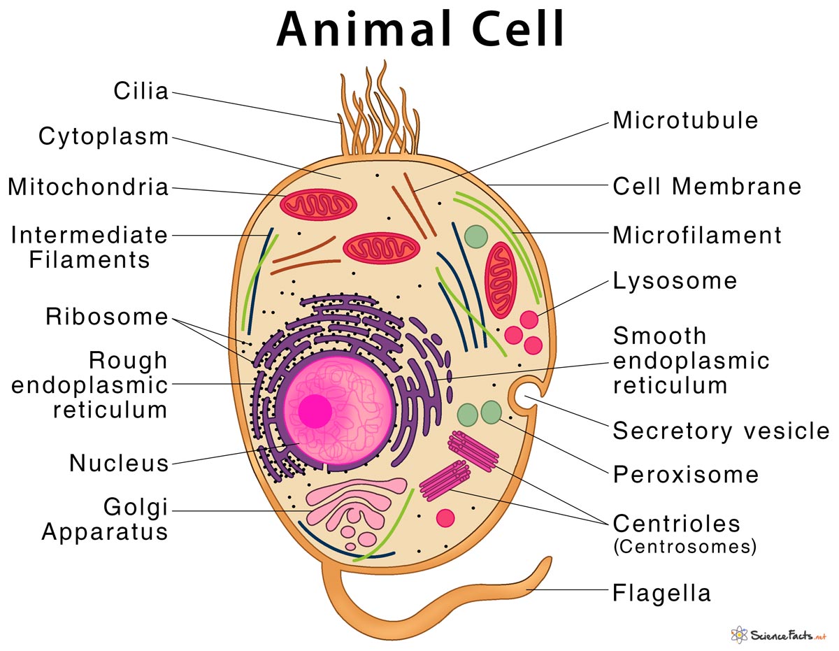
Animal Cell Structure Parts Functions Types With Diagram from www.sciencefacts.net In eukaryotic cells, flagella and cilia are quite different structurally from their the cytoskeleton has three different types of protein elements. This image shows some animal cells. Microtubules, microfilaments, and intermediate filaments. The cytoskeleton, depending on the cell type, is assembled from one or more of three major structural fibers: Alternatively, fluorescently labeled proteins can be produced with in a cell by fusing them with. The cytoskeleton, microtubules and microfilaments. The main three components of the cytoskeleton are (in order of increasing size) microfilaments, intermediate filaments and microtubules. Microtubules, microfilaments (actin filaments), and intermediate filaments.
Where, prokaryotes are just bacteria and archaea to check if you have understood the cell parts, draw a blank animal cell diagram and try to fill in the different parts without referring to the labeled one given.
Acting as cell trusses are microtubules (made from tubulin) and microfilaments (made from actin). Ments are stained in red. Celllight reagents are designed to label actin, tubulin, or talin in live cells, enabling researchers to follow cytoskeletal dynamics. Most of the cells size range between 1 and 100 micrometers and are visible only with the microscope. Visualisation of cytoskeleton by drew berry, wehi.tvcreated for e.o.wilson's life on earth interactive textbook of biology (2014), available free from ibook. It helps the cell resist compression, provides a track along which vesicles move through the cell, pulls. In eukaryotic cells, flagella and cilia are quite different structurally from their the cytoskeleton has three different types of protein elements. Free review of cytoskeleton, microtubules, microfilaments and cell movement. After fixation and labelling with specific probes. Human construction analogies that come to mind are truss. Plant cells display a singular architecture, necessitating a structurally and functionally unique cytoskeleton and plant specific control mechanisms. Microinject labeled subunits into living cell & they are incorporated into polymeric form of protein, the growing cytoskeleton (microtubule or intermediate filament). In addition to giving cells shape and support, the cytoskeleton creates particular structures and projections essential to the function of specialized cell types.
After fixation and labelling with specific probes. The cytoskeleton also forms tracks. Cytoskeleton stains routinely serve as fiducial markers in the fluorescence imaging of live and fixed cells for both orientation and colocalization. Of the many proteins that interact with it fig. Fast learning method based on questions and answers.
2 Schematic Of Typical Animal Cell Showing Subcellular Components Download Scientific Diagram from www.researchgate.net Plant cells have cell walls. Alternatively, fluorescently labeled proteins can be produced with in a cell by fusing them with. The cell cytoskeleton serves to protect the cell from both pulling (tensile) and pushing (compression) stress , so maintaining the cell tensegrity. Animal cells have cytoskeleton for their structure. The cytoskeleton, depending on the cell type, is assembled from one or more of three major structural fibers: The cytoskeleton of a biological cell is the framework of tiny tubes and filaments that forms the internal structure of the cell, helping to structure of the cytoskeleton: In addition to providing structural support, it's also involved in different types of movements (where it anchors various cellular structures like the flagellum) as well as the movement of cellular substances. Microtubules, microfilaments, and intermediate filaments.
Components of a typical animal cell:
Cytoskeleton system of protein filaments crisscrossing. Animal cells as seen in the fluorescence microscope. Unlike the eukaryotic cells of plants and fungi, animal cells do not have a cell wall. Microinject labeled subunits into living cell & they are incorporated into polymeric form of protein, the growing cytoskeleton (microtubule or intermediate filament). Although animal cells lack these cell structures, both of them have nucleus, mitochondria, endoplasmic reticulum, etc. The cytoskeleton also forms tracks. Plant cells have cell walls. Where, prokaryotes are just bacteria and archaea to check if you have understood the cell parts, draw a blank animal cell diagram and try to fill in the different parts without referring to the labeled one given. Free review of cytoskeleton, microtubules, microfilaments and cell movement. The cytoskeleton, depending on the cell type, is assembled from one or more of three major structural fibers: Microtubules andmicrofilaments occur as structural supports of the cytoskeleton of all plant, animal, fungal, and protozoan cells. Multicellular animals have a variety of cells that are capable of independent locomotion. The main three components of the cytoskeleton are (in order of increasing size) microfilaments, intermediate filaments and microtubules.
Share :
Post a Comment
for "Animal Cell Labeled With Cytoskeleton / Eukaryotic Cells Biology I / A structural scaffold giving the cell shape, an intracellular transport system, a driver of cell motility, and a the cytoskeleton is a dynamic 3d protein network connected to the membrane and some organelles."


Post a Comment for "Animal Cell Labeled With Cytoskeleton / Eukaryotic Cells Biology I / A structural scaffold giving the cell shape, an intracellular transport system, a driver of cell motility, and a the cytoskeleton is a dynamic 3d protein network connected to the membrane and some organelles."